
Endoscopy in Nashik
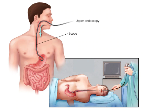
Upper endoscopy enables visual examination of the upper digestive system using a tiny camera mounted on one end of a long and flexible tube. Endoscopy is used by a physician specializing in diseases of the digestive system (gastroenterologist) to diagnose and occasionally treat conditions affecting the esophagus, stomach, and first part of the small intestine. An alternative name for an upper endoscopy is esophagogastroduodenoscopy an expert examines the esophagus, stomach as well as the start of the small intestine to determine if the patient is suffering from a disease that attacks the digestive system through the use of an endoscope. The disease that attacks the digestive system is usually diagnosed using endoscopy techniques, especially for conditions such as ulcers or tumours within the gastrointestinal tract. In case the patient wants such a procedure done on the patient visit the patient’s physician for an upper endoscopy.”
How patient prepare
Fast before the endoscopy. The patient will typically need to stop eating solid food for eight hours and stop drinking liquids for four hours before the patient’s endoscopy. This is to ensure the patient’s stomach is empty for the procedure.
Stop taking certain medications. The patient will need to stop taking certain blood-thinning medications in the days before the patient’s endoscopy, if possible. Blood thinners may increase a patient’s risk of bleeding if certain procedures are performed during the endoscopy. If a patient has ongoing conditions, such as diabetes, heart disease or high blood pressure, the patient’s provider will give the patient specific instructions regarding the patient’s medications.
Plan for the patient’s recovery!
Most people undergoing an upper endoscopy will receive a sedative to relax them and make them more comfortable during the procedure. Plan for the patient’s recovery while the sedative wears off. The patient may feel mentally alert, but the patient’s memory, reaction times and judgment may be impaired. Find someone to drive the patient home. Patients may also need to take the day off from work. Don’t drive or make any important personal or financial decisions for 24 hours.
What patients can expect during an endoscopy?
Monitors often are attached to the patient’s body. This allows the patient’s healthcare team to monitor the patient’s breathing, blood pressure and heart rate.
The patient may receive a sedative medication. This medication, given through a vein in the patient’s forearm, helps the patient relax during the endoscopy.
An anaesthetic may be used in the patient’s mouth. An anaesthetic spray numbs the patient’s throat in preparation for the insertion of the long, flexible tube (endoscope). The patient may be asked to wear a plastic mouth guard to hold the patient’s mouth open.
Then the endoscope is inserted in the patient’s mouth. The patient’s provider may ask the patient to swallow as the scope passes down the patient’s throat. The patient may feel some pressure in the patient’s throat, but the patient shouldn’t feel pain.
The patient can’t talk after the endoscope passes down the patient’s throat, though the patient can make noises. The endoscope doesn’t interfere with the patient’s breathing.
A tiny camera at the tip transmits images to a video monitor in the exam room. The patient’s provider watches this monitor to look for anything out of the ordinary in the patient’s upper digestive tract. If something unusual is found in the patient’s digestive tract, images can be taken for later examination.
Gentle air pressure may be fed into the patient’s esophagus to inflate the patient’s digestive tract. This allows the endoscope to move freely. It allows the folds of the patient’s digestive tract to be examined more easily. The patient may feel pressure or fullness from the added air.
The patient’s provider will pass special surgical tools through the endoscope to collect a tissue sample or remove a polyp. This is done with the help of a video monitor to guide the tools.
Risks Factors
Bleeding. A patient’s risk of bleeding complications after an endoscopy is increased if the procedure involves removing a piece of tissue for testing (biopsy) or treating a digestive system problem. In rare cases, such bleeding may require a blood transfusion.
Infection. Most endoscopies consist of an examination and biopsy, and the risk of infection is low. The risk of infection increases when additional procedures are performed as part of the patient’s endoscopy. Most infections are minor and can be treated with antibiotics. The patient’s doctor may give the patient preventive antibiotics before the patient’s procedure if the patient is at a higher risk of infection.
Tearing of the gastrointestinal tract. A tear in the patient’s esophagus or another part of the patient’s upper digestive tract may require hospitalization, and sometimes surgery to repair it. The risk of this complication is very low, but it increases if additional procedures, such as dilation to widen the patient’s esophagus, are performed.
Reaction to sedation. Before the patient’s upper endoscopy, the patient will likely be given sedation so that the patient will be better able to tolerate the procedure. The type of sedation varies, and adverse reactions are possible but rare. Patients will be monitored closely during the procedure to reduce the risk of a serious reaction.
Laparoscopic Surgery In Nashik?
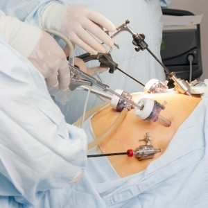
Laparoscopy, also known as diagnostic laparoscopy, is a surgical diagnostic procedure used to examine the organs inside the abdomen. It’s a low-risk, minimally invasive procedure that requires only small incisions.
Laparoscopy uses an instrument called a laparoscope to look at the abdominal organs. A laparoscope is a long, thin tube with high-intensity light and a high-resolution camera at the front. The instrument is inserted through an incision in the abdominal wall. As it moves along, the camera sends images to a video monitor.
Laparoscopy allows the patient’s doctor to see inside the patient’s body in real-time, without open surgery. Patient doctors also can obtain biopsy samples during this procedure.
Why is Laparoscopy performed?
- Ultrasound, which uses high-frequency sound waves to create images of the body.
- CT scan, which is a series of special X-rays that take cross-sectional images of the body.
- MRI scan, which uses magnets and radio waves to produce images of the body.
What are the risks of Laparoscopy?
- Fevers or chills
- Abdominal pain that becomes more intense over time.
- Redness, swelling, bleeding, or drainage at the incision sites.
- Continuous nausea or vomiting.
- Persistent cough.
- Shortness of breath.
- Inability to urinate.
- Light-headedness